
Figure 6.
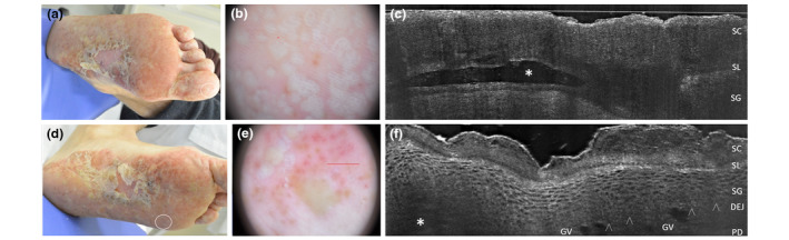
Palmoplantar pustular psoriasis of acute onset in a 38‐year‐old man. Clinical, dermoscopic and LC‐OCT appearance of early developed lesions of finger toe (a), visible as white‐yellowish roundish structure under dermoscopy (b) and as an upper‐epidermal area with well‐defined borders and hyper‐reflective roundish structures corresponding to neutrophils in LC‐OCT (c, asterisk). The LC‐OCT examination carried out at plantar perilesional site (d, white circle) performed near a plantar pustule (f, asterisk) demonstrates the presence of well‐defined large capillary loops in the papillary dermis (f), corresponding to the red clots (e) visible in the dermoscopic image [SC, stratum corneum; SL, stratum lucidum; SG, stratum granulosum; DEJ: dermo‐epidermal junction; arrowheads: DEJ profile; CV: capillary vessels; harrowheads: dermo‐epidermal junction. The red line inside polarized dermoscopy 159 (b) corresponds to the LC‐OCT vertical frame].