Figure 7.

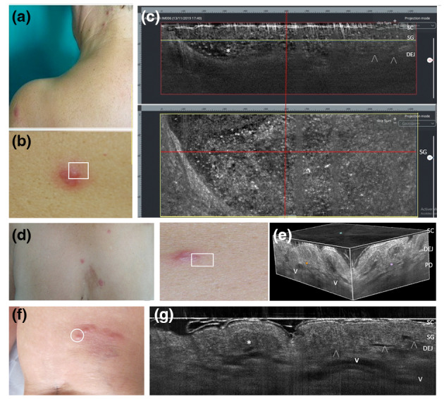
Sweet syndrome with vesicopustular appearance and multifocal lesion distribution in two women aged 62 (a–c) and 55 years (d–e). Combined vertical and horizontal LC‐OCT frames (c) of a scapular lesion (b) revealed dense roundish a‐reflective area with well‐defined borders filled with multiple floating hyper‐reflective roundish structures (c, asterisks), corresponding to neutrophilic collections in the epidermis and upper dermis (d); 3D LC‐OCT of a chest lesion (d) showed the distribution of dilated vessels (e, V) and the presence of papillary oedema as hypo‐reflective spaces in the dermis. Differential diagnosis with a case of eosinophilic cellulitis with papulo‐pustular appearance in a 58‐year‐old woman (f), where LC‐OCT shows an intraepidermal area with dense non‐homogenous content in the epidermis/DEG (i, asterisk) corresponding to a non‐homogeneous collection of eosinophils and neutrophils, overlying dilated tortuous vessels (g, V) in the whole dermis. [SC, stratum corneum; SL, stratum lucidum; SG, stratum granulosum; DEJ: dermo‐epidermal junction; arrowheads: DEJ profile; V: vessels. The horizontal yellow line in the vertical LC‐OCT frame (c, red box) indicate the depth level of the horizontal frame (c, yellow box) within the SG].
