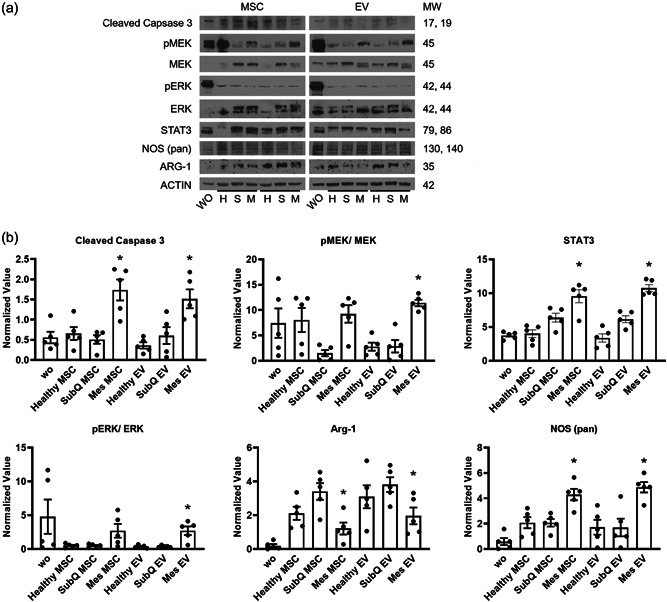
Figure 6.

Enhanced cleaved caspase 3, MEK, ERK, STAT3, and suppressed Arg‐1 signaling in inflamed extracellular vesicles incubated macrophages. THP‐1 cells were exposed to inflamed, subcutaneous or noninflamed MSCs or extracellular vesicles for 72 h in macrophage differentiation conditions before protein harvest and western blot analysis. (a) The levels of cleaved caspase 3, pMEK, MEK, pERK, ERK, STAT3, and ARG‐1 after MSCs treatment were determined by immunoblotting. (b) The levels of cleaved caspase 3, pMEK, MEK, pERK, ERK, STAT3, and ARG‐1 after extracellular vesicles treatment were determined by immunoblotting. Relative band intensities were quantified from five individual experiments and measured by densitometry analysis. Beta‐actin was used as a loading control. Phosphorylated proteins were further normalized by total protein values. Mes, diseased mesentery; MSC, mesenchymal stem cells; Subq, subcutaneous; WO, without treatment. n = 3 patients. *p < 0.05.
