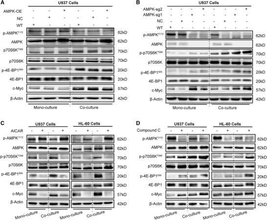
FIGURE 5.

AMPK inhibition increases mTORC1 activity and c‐Myc proteins expression. (A) Representative western blot analysis of AMPK‐mTORC1 pathway proteins (total AMPK, p‐AMPKT172, total p70S6K, p‐p70S6KT389, total 4E‐BP1, p‐4E‐BP1S65, and c‐Myc; β‐actin was used as loading control) of AMPK‐overexpressing U937 cells. (B) Representative western blot analysis of AMPK‐mTORC1 pathway proteins in U937 cells knocked out for AMPK expression. (C) Representative western blot analysis of AMPK‐mTORC1 pathway proteins from AML cells after treatment with 1 mM AICAR. (D) Representative western blot analysis of AMPK‐mTORC1 pathway proteins in AML cells after treatment with 5 μM Compound C. WT, wide type; NC, empty vector control; AMPK‐OE, AMPK overexpression; AMPK‐sg1/2, AMPK gene was knocked out. The representative images of 3 independent experiments were used
