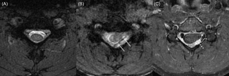
FIGURE 3.

Axial cervical MRI on the neutral (A) and flexion position (B and C). Spoiled T2*‐weighted image at the level of C5 in neutral position shows a normal spinal cord morphology (A). Spoiled T2*‐weighted (B) and post‐contrast T1‐weighted (C) images at the level of C5 reveal the forward displacement of the posterior dural sac (arrows in B) with asymmetrical compression of the spinal cord, more prominent on the left side (dashed arrow in C)
