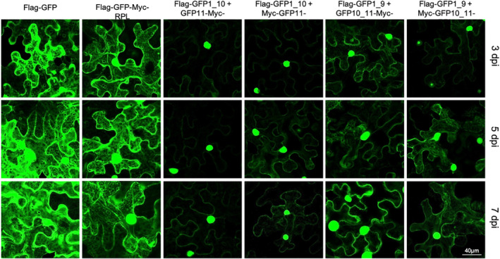
Figure 2.

Comparison of fluorescence from green fluorescent protein (GFP) protein splits and different tag positions. Confocal microscopy was used to assay GFP activity and subcellular localization in Nicotiana benthamiana leaves transiently transformed through infiltration of Agrobacterium containing the genes of interest in T‐DNA vectors. Images were recorded as z‐stacks of multiple planes. dpi, days post‐infiltration with Agrobacterium; labels correspond to the constructs that are illustrated in Figure 1(c). [Colour figure can be viewed at wileyonlinelibrary.com]
