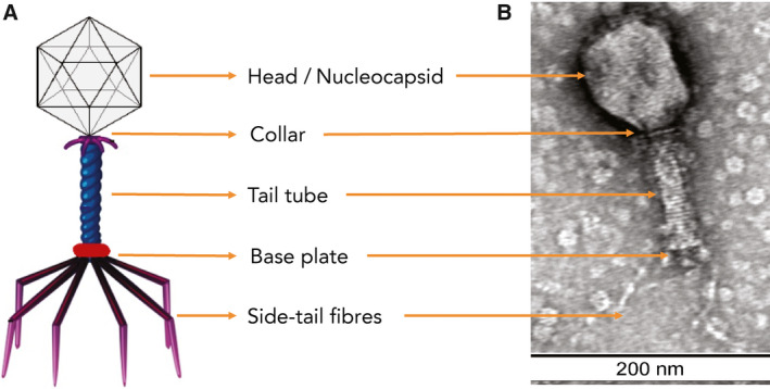
Schematic (A) and electron micrograph of bacteriophage (Myoviridae) with 1% uranyl acetate negative staining, with size marker (B). Bacteriophage preparations were dialysed against 0.1 M ammonium acetate in dialysis cassettes with a 10 000 membrane molecular weight cut‐off (Pierce Biotechnology), negatively stained with 2% uranyl acetate, and visualised using transmission electron microscopy (TEM). TEM was conducted at the Westmead Scientific Platforms (Westmead Hospital, Sydney, Australia) on a Philips CM120 BioTWIN (Thermo Fisher Scientific) transmission electron microscope at 100 kV. Images were recorded with a SIS Morada digital camera using iTEM software (Olympus Soft Imaging Solutions).
