Fig. 3.

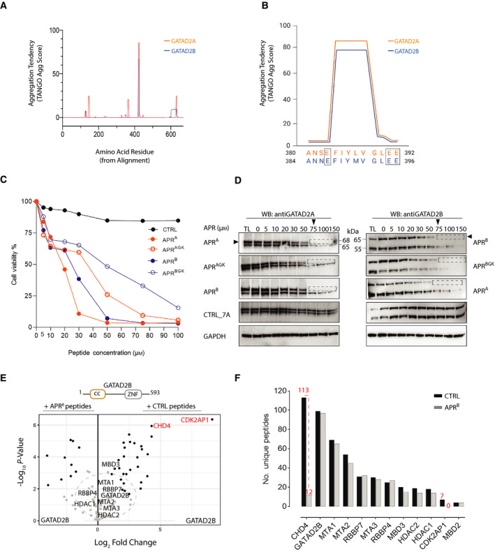
An APR within GATAD2 proteins co‐ordinates CDK2AP1 and CHD4 association with NuRD. (A) TANGO analysis shows β‐sheet aggregation tendency for the APR within GATAD2A and GATAD2B (B) A close‐up schematic of the β‐sheet aggregation tendency of the APRs in GATAD2A (384‐390) and GATAD2B (388‐394) and gatekeeper residues (boxed). (C) Graph representing the MTT assay performed in 2 biological and three technical replicates (n = 6). Cell viability of K562 cells treated with a range of APR peptide concentrations was measured after 48 h. Data are presented as a per cent of untreated cell viability measured using the MTT assay. (D) Western blots of endogenous GATAD2 proteins after treatment of K562 cell lysates with APR‐tat peptides. APR peptidomimetics (0–150 μm) were added to the lysates before sonication. Total lysates (TL, 5%) were taken as input before separation of the soluble and insoluble fractions by centrifugation. Cleared lysate (5% of soluble fraction) was run on SDS‐PAGE and probed for GATAD2 proteins. Black arrows indicate the canonical isoform of GATAD2A and GATAD2B proteins, and dotted rectangular box shows loss of soluble proteins post‐APR treatment. (E) Volcano plot comparing the interactors of overexpressed GATAD2B in the presence of 50 μm APRB (left panel) or CTRL (right panel) peptides. (F) The number of unique peptides of each subunit of the NuRD complex detected by MS after FLAG‐GATAD2B PD‐MS in the presence of APRB and CTRL peptides.
