
FIGURE 2.
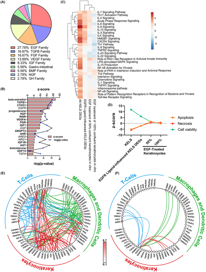
Growth factor signalling is altered in atopic dermatitis lesional skin. (A‐B) Growth factor upstream mediators grouped by family in (A) and the most significantly enriched growth factors and hormones given in (B). (C) Inflammatory pathways enriched in differently expressed genes (DEGs) between keratinocyte‐enriched lesional (KELS), KELS DEGs downstream of EGFR ligands and non‐lesional skin and in EGF‐treated human keratinocytes. 36 (D) Cell viability/death pathways in KELS, KELS DEGs downstream of EGFR ligands and in EGF‐treated human keratinocytes. 36 (E,F) Ligand–receptor interaction plots between in situ keratinocytes, T cells and macrophages/dendritic cells for all (E) growth factors or (F) EGF family ligands in AD lesional skin. Ligands (circles) and receptors (squares) are coloured by relative expression in lesional skin. DEGs are considered p < 0.05 using a one‐sample t‐test with multiple testing correction using the BH method. Heatmaps use Euclidean distance and complete linkage