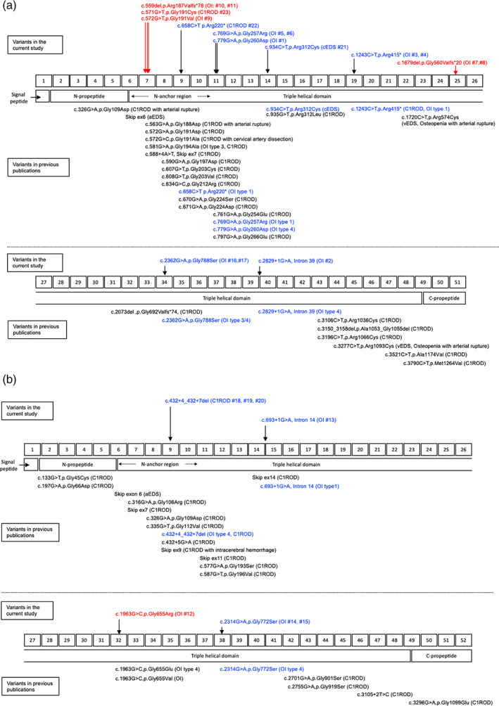
FIGURE 1.

Schematic structure, location of domain organizations, and distribution of the mutations in COL1A1 (a) and COL1A2 (b). The numbers in the upper center rectangles indicate the exon numbers, and the rectangles in the lower center indicate the domain organizations of the proteins. Variants found in the current study are shown above the exon rectangles; unpublished variants are displayed in red, and previously published variants are displayed in blue. Variants in previous publications are shown below the domain rectangles. Variants both identified in the current study and previous publications are displayed in blue. aEDS, Ehlers‐Danlos syndrome arthrochalasia type; C1ROD, COL1‐related overlap disorder; OI, osteogenesis imperfecta; cEDS Ehlers‐Danlos syndrome classical type; vEDS, Ehlers‐Danlos syndrome vascular type
