
Fig. 2.
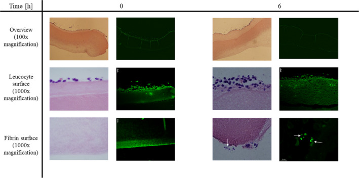
Biofilm development is microscopically visible after 6 h. IEVs were inoculated with 5 × 10⁴ CFU GFP‐tagged Staphylococcus aureus and 2 IEVs were transferred into 4% formalin either right after infection (0 h), or after 3, 6, and 9 h. Fixated IEVs were stored in formalin in the fridge until usage. IEVs were then embedded in paraffin, and vertical slices were prepared for microscopy by H&E staining (light microscopy, left panels) or deparaffination for detection of GFP‐tagged bacteria (fluorescence microscopy, right panels) to ensure that the observed aggregates are indeed S. aureus. Displayed are representative areas of 2 biopsies at 0 and 6 h after inoculation at 100‐ and 1000‐fold (H&E) or 630‐fold (GFP) magnification. Biofilm‐like bacterial aggregates (white arrows) were observed after 6 and 9 (data not shown) hours and located exclusively on the fibrin‐surface of the IEV.