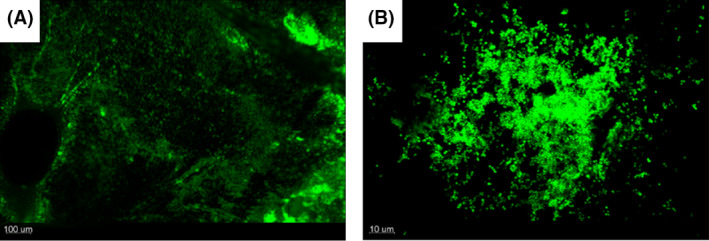
Fig. 3.

Confocal laser scanning microscopy reveals uneven distribution of biofilms on the fibrin surface of the IEV. IEVs were inoculated with 5 × 10⁴ CFU GFP‐tagged Staphylococcus aureus and incubated for 24 h. IEVs were washed twice in 200 μL saline and loaded onto microscopy slides with the fibrin surface facing up. Bacterial biofilm formation was observed with confocal laser scanning microscopy in 100x magnification (A) and 630× magnification (B). Displayed is the top‐down view of a representative area of one IEV (A) and one of the aggregates in close up (B).
