Figure 1.

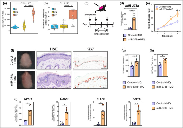
miR‐378a is upregulated in keratinocytes in human lesional psoriatic skin and increases skin thickness through promoting inflammation and keratinocyte hyperproliferation in mice. CD45− epidermal cells (mainly keratinocytes) were separated from skin biopsies obtained from lesional (PP) and nonlesional (PN) skin of patients with psoriasis (n = 9), and from healthy (H) controls (n = 9) by magnetic cell sorting. (a) Read count (transcripts per million) of miR‐378a in keratinocytes (epidermal CD45− cells) from H, PN and PP skin. Data are extracted from small RNA sequencing. (b) Reverse‐transcription quantitative real‐time polymerase change reaction (RT‐qPCR) analysis of miR‐378a in keratinocytes from PP and PN skin (n = 20), and from H skin (n = 19). Expression is normalized to U48 RNA and shown as relative expression units. (c) Timeline for topical application of imiquimod (IMQ) and intradermal (ID) injection of miR‐378a mimics or control oligos (arrows) on mouse back skin. (d) RT‐qPCR analysis of miR‐378a levels in mouse skin upon ID delivery of miR‐378a mimics. (e) Changes in skin thickness upon induction of psoriasis‐like skin inflammation by the topical application of IMQ on mice injected with miR‐378a mimic (orange) or control oligos (scramble mimic, purple). (f) Representative macroscopic imaging (left), haematoxylin and eosin (H&E) staining (centre) and immunohistochemistry for Ki67 protein (right) of back skin sections from mice treated with miR‐378a mimic or control oligo in combination with IMQ application. (g) Quantification of the epidermal thickness on H&E staining of back skin. (h) Quantification of Ki67 positive cells in the epidermis of back skin. (i) RT‐qPCR analysis of Cxcl1, Ccl20, Il‐17c and Krt16 in back skin samples. Scale bars = 50 μm. *P < 0·05, **P < 0·01, and ***P < 0·001, ****P < 0·0001; n = 6 per group. Mann–Whitney U‐test was used. [Colour figure can be viewed at wileyonlinelibrary.com]
