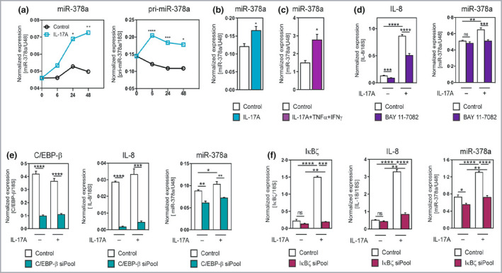
Figure 2.

Interleukin (IL)‐17A induces miR‐378a expression in primary keratinocytes through nuclear factor kappa B (NF‐κB) and C/EBP‐β. (a) Primary human epidermal keratinocytes were treated with the cytokine IL‐17A at a concentration of 100 ng mL−1 for the indicated times; the primary transcript pri‐miR‐378a and mature miR‐378a expression was analysed by reverse‐transcription quantitative real‐time polymerase change reaction (RT‐qPCR). Expression is normalized to 18S for the primary transcript and U48 RNA for the mature miRNA and data points show the mean of four replicates. (b) RT‐qPCR analysis of miR‐378a expression in three‐dimensional (3D) epidermal equivalents treated with IL‐17A (20 ng mL−1) for 72 h. (c) RT‐qPCR analysis of miR‐378a expression in 3D epidermal equivalent treated for 72 h with a combination of cytokines associated with psoriasis (psoriasis‐like), i.e. IL‐17A (30 ng mL−1) + tumour necrosis factor (TNF)‐α (5 ng mL−1) + interferon (IFN)‐γ (20 ng mL−1). (d) RT‐qPCR analysis of miR‐378a expression in primary human keratinocytes treated with IL‐17A (100 ng mL−1) with or without pretreatment with the NF‐κB inhibitor BAY11‐7082. The expression of miR‐378a was normalized to U48 RNA. Bars show mean ± SD of four replicates. (e) RT‐qPCR analysis of miR‐378a and IL‐8 expression in keratinocytes upon inhibition of C/EBP‐β by small interfering RNA (siRNA) pool. (f) RT‐qPCR analysis of miR‐378a and IL‐8 expression in keratinocytes upon inhibition of IκBζ by an siRNA pool. *P < 0·05, **P < 0·01, ***P < 0·001, ****P < 0·0001, ns: not significant, Student’s two‐sided t‐test was used. [Colour figure can be viewed at wileyonlinelibrary.com]
