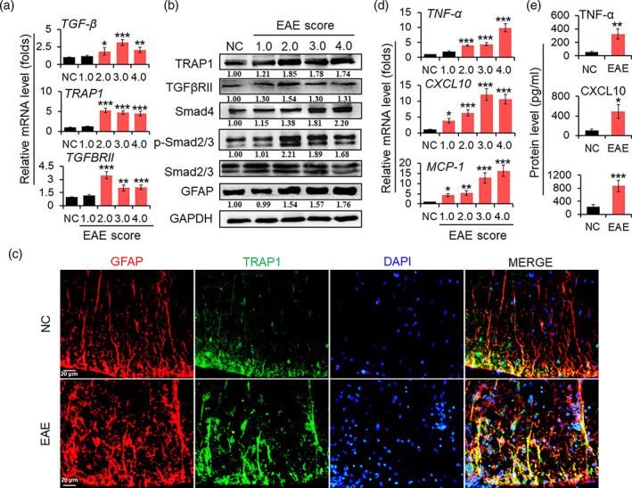
FIGURE 1.

TRAP1/Smad signal pathway is activated in EAE mice. (a) The transcriptional level of TGF‐β, TRAP1, and TGFβRII mRNAs in spinal cords of EAE mice was tested by real‐time PCR assay. (b) The protein level of TRAP1, TGFβRII, Smad4, p‐Smad2/3, and GFAP in spinal cords was measured by Western blot assay. The relative level TRAP1, TGFβRII, Smad4, and GFAP was determined by quantitative densitometry compared to GAPDH; the relative level of phosphorylation Smad2/3 (p‐Smad2/3) was determined by quantitative densitometry after normalized to corresponding non‐phosphorylation ones. The relative value of proteins in the NC group was considered to be 1 for comparison. (c) IFA was used to test the expression of GFAP (red), TRAP1 (green), and nuclear staining of DAPI (blue) in astrocytes from spinal cords of EAE mice. Scale bars, 20 μm. (d) The mRNA levels of TNF‐α, CXCL10, and MCP‐1 in spinal cords were determined by real‐time PCR assay. (e) The secretion levels of TNF‐α, CXCL10, and MCP‐1 in the sera of mice were detected by ELISA. Data are represented as the mean ± SEM. *p < .05, **p < .01, and ***p < .001 versus NC group (n = 10 mice/group)
