Figure 1.

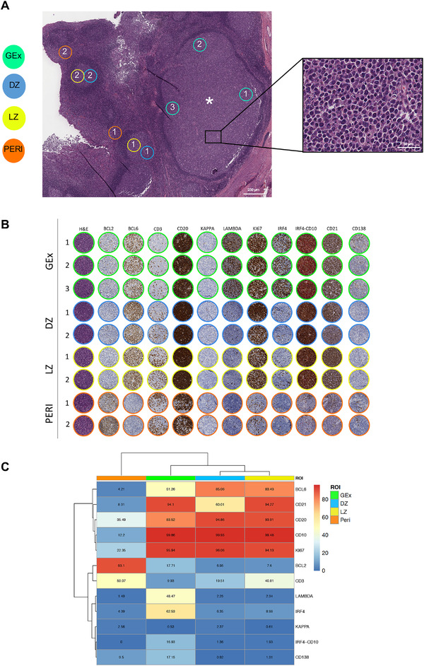
(A) Digitalized slide selection of the H&E‐stained section of the tonsil highlighting the presence of an aberrantly expanded GC (asterisk) characterized by the presence of elements with plasmacytoid morphology (inset). On the H&E, representative regions relative to GC dark zone (DZ) and light zone (LZ) areas, perifollicular (Peri) areas, and germinotropic plasmablastic expansion (GEx) areas, are highlighted. Original magnification ×50. (B) Comparative analysis of H&E and IHC for Bcl‐2, Bcl‐6, CD3, CD20, Kappa and Lambda light chain, Ki67, IRF4, IRF4/CD10, CD2, CD138 in the DZ, LZ, Peri, and GEx areas highlighted in (A). (C) Heatmap of the average expression of the quantitative immunohistochemical analysis of the markers evaluated in the DZ (n = 5), LZ (n = 5), Peri (n = 5), and GEx (n = 9) areas highlighted in (B).
