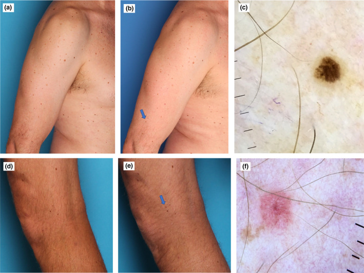
Figure 2.

(a–c) Man in his 50s with dysplastic naevus syndrome. (a,b) A tiny melanoma 1.5 mm in diameter in situ detected on the comparison of the total body photographs (blue arrow) at (a) baseline and (b) at the 12‐month follow‐up, which revealed a completely new pigmented lesion. (c) Cross‐polarized dermoscopy (MoleScope II; MetaOptima Technology) findings were subtle, showing two colours and a few irregular peripheral brown dots (original magnification × 10). (d–f) Man in his 40s with a personal history of melanoma and dysplastic naevus syndrome. (d,e) Tiny invasive amelanotic melanoma (2.5 mm in diameter, Breslow thickness 0.2 mm, no ulceration, no mitoses) detected as a new lesion (blue arrow) on comparison of total body photographs at (d) baseline and (e) 12‐month follow‐up. (f) Cross‐polarized dermoscopy (Dermmlite DL4; 3Gen, Orange County, CA, USA) showed shiny white lines and dots and atypical vascular pattern with dotted vessels (cross‐polarized dermoscopy, original magnification × 10). [Colour figure can be viewed at wileyonlinelibrary.com]
