FIGURE 3.

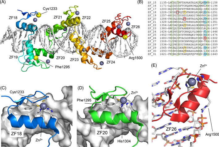
Structural model of the missense variants of ZNF142. (A) Structural model of ZF motifs 18–26 (ZF18 ‐ ZF26) of the protein and their interaction with DNA. Zn2+ ions are represented as gray spheres. The positions of the residues Cys1233, Phe1295, and Arg1500 are indicated. (B) Multiple sequence alignment of the 17 C‐terminal ZF motifs. The positions of Cys1233 (ZF18), Phe1295 (ZF20), and Arg1500 (ZF26) are shown in red. In green, the Cys and His residues that coordinate the Zn2+ ion (among them, Cys1233). In yellow, positions homologous to Phe1295 that are conserved. In blue, conserved positions with positive electrostatic charge homologous to Arg1500. (C) Position of Cys1233 coordinating the Zn2+ ion. The Cys1233Phe substitution would completely disorder the structure of ZF18. (D) Position of Phe1295 stabilizing the position of His1304, which, in turn, coordinates the Zn2+ ion. (E) Position of Arg1500 associating with the DNA phosphate chain. The Arg1500Trp substitution would prevent the correct interaction of ZF26 with DNA [Colour figure can be viewed at wileyonlinelibrary.com]
