
FIGURE 3.
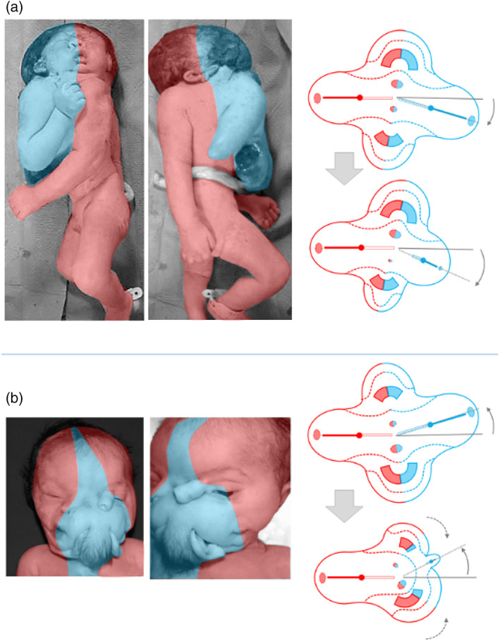
Asymmetric cephalothoracoileopagus. (a) Case described by Kaufmann (Kaufmann et al., 2007) including schematics of the embryonic disk model before (top) and after parasitic regression (bottom). Source: Reprinted with permission from TheFetus.net. (b) Case described by Kastenbaum (Kastenbaum, McPherson, Murdoch, & Ozolek, 2009) including schematics of the embryonic disk model before (top) and after parasitic regression (bottom). As a result of the profound regression, the neo‐axial orientation of compound structures and organs in the convex and concave conjunction planes is largely abolished (dashed arrows). Source: Used with permission from Sage journals