
FIGURE 1.
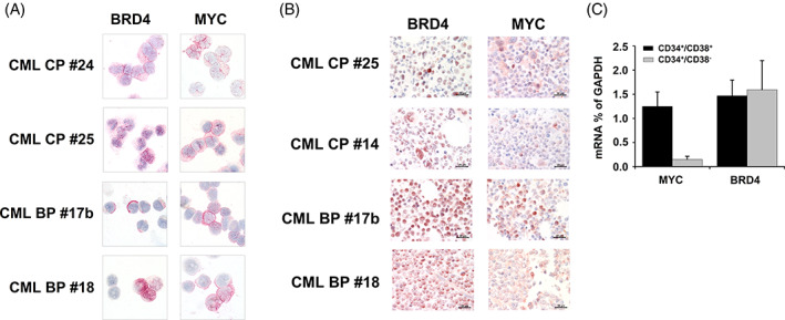
Expression of BRD4 and MYC in CML cells. (A) Immunocytochemical evaluation of BRD4 and MYC expression in primary CML MNC isolated from 2 patients with CML in chronic phase (CP) and 2 patients with CML blast phase (BP) was performed using a polyclonal antibody against BRD4 and a monoclonal antibody directed against MYC. Original magnification, ×100. (B) Immunohistochemical detection of BRD4 (left panels) and MYC (right panels) in CML in bone marrow biopsy sections in 2 patients with CML CP and 2 patients with CML BP. Original magnification, ×60. Slides were investigated using an Olympus DP21 camera connected to an Olympus BX50F4 microscope equipped with ×60/0.90 UPlanFL (IHC) or ×100/1.35 UPlanAPO (Oil Iris; ICC) objective lenses. Images were adjusted by Adobe Photoshop CS5. C: qPCR was performed using sorted CD34+/CD38+ and CD34+/CD38− cells from patients with CP CML (n = 3). Results are expressed as BRD4 and MYC mRNA levels relative to (as percent of) GAPDH mRNA levels and represent the mean ± SD from three independent experiments. [Color figure can be viewed at wileyonlinelibrary.com]