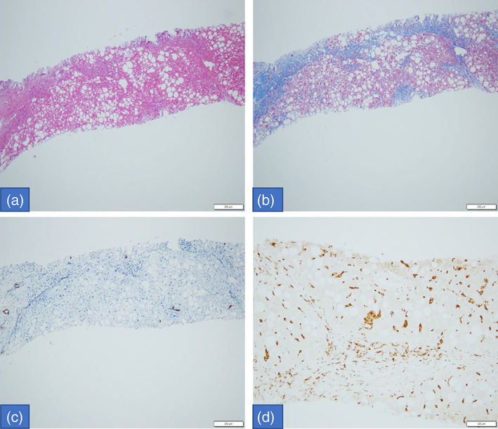
FIGURE 2.

Histologic exam of liver biopsy: macrovesicular steatosis with few portal‐inflammatory cells (HE. 10×); disturbed architecture due to porto‐portal fibrous septa (Masson trichrome stain, 10×); loss of biliary ducts (CK7 IHC, 10×) and increased Kupffer cells, sometimes with large leaf‐like cytoplasm (CD68 IHC, 20×).
