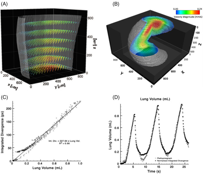
FIGURE 4.

Preclinical experiments validating the accuracy and validity of lung volume measures using XV technology, with (A) and (B) showing bench‐top measurements of fluid flow validated against computer modelling; and (C) and (D) showing in vivo measurements of ventilation in rabbit lungs validated against plethysmography. (A) Reconstructed 3D blood velocity flow fields measured using XV. For clarity only half the sample is plotted, with reduced vector resolution in all dimensions. Vector colours represent velocity magnitude and are validated against computational models of the flow field. (B) CT XV reconstruction of flow field through helical geometry. A section of the result has been rendered as transparent for visualization of the flow. The results indicate the ability of CT XV to simultaneously measure the 3D structure and velocity of flow through complex geometries. (C) In vivo measurements of ventilation in rabbit lungs with validation of integrated divergence (volume) measurements from XV technology against volume measures from plethysmography. A scatter plot shows strong correlation between two quantities. (D) Time series of lung volume co‐plotted with divergence demonstrated a direct link between divergence and tissue expansion
