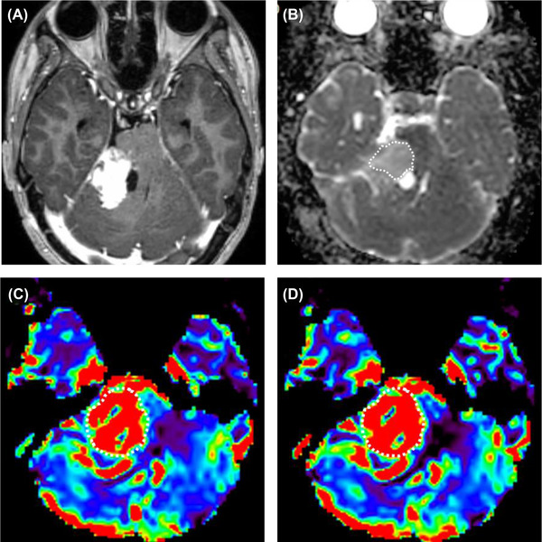
FIGURE 3.

An 18‐year‐old neurofibromatosis type 2 female with schwannoma in the right cerebellopontine angle. (A) Postcontrast T1‐weighted image shows a heterogeneously enhancing mass with a cystic component in the medial aspect, in the right cerebellopontine angle. (B) The ADC map was generated, and the calculated normalized ADC mean was 1.34. (C) The rCBV and (D) rCBF were generated from DSC‐MRI, and calculated nrCBV and nrCBF were 5.05 and 5.83, respectively. nrCBV, normalized relative cerebral blood volume; nrCBF, normalized relative cerebral blood flow.
