Figure 2.

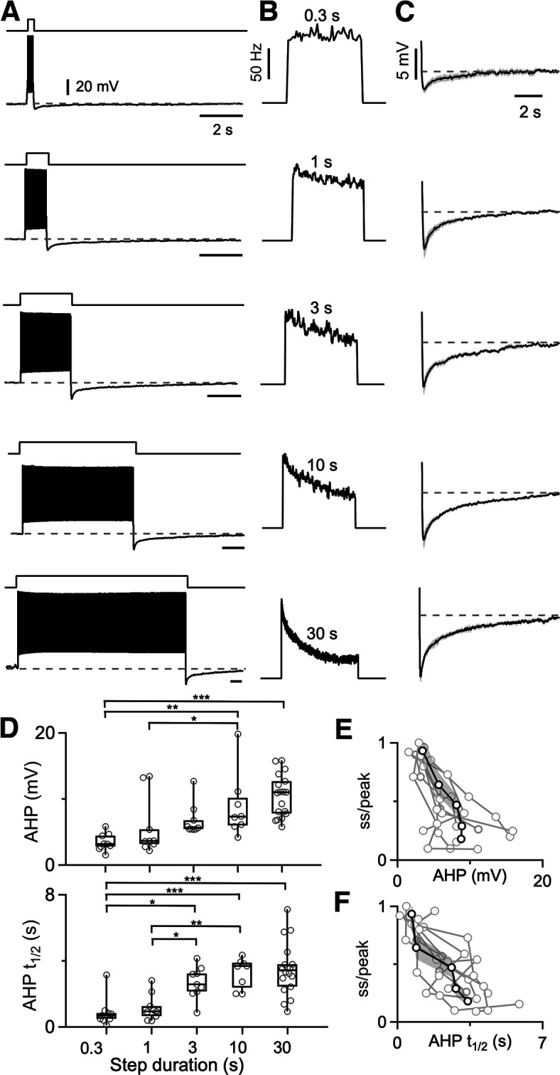
Prolonged spiking leads to a slow afterhyperpolarization that is dependent on spiking duration. A, Firing and AHP for a DCN neuron are shown in response to delivering depolarizing current steps of varying duration. B, Instantaneous firing frequencies are shown for the spiking shown in A. C, Voltage traces showing the average AHP following 0.3, 1, 3, 10, and 30 s current steps (N = 7 cells from 5 animals; SEs are in gray). D, Summaries of the amplitudes and half-recovery times of the AHPs for individual cells (circles) after current steps of the indicated durations. Half-recovery time was calculated as half the time for the AHP to return to the initial membrane potential before the step. N = 7 cells (5 animals). One-way ANOVA was performed to compare the AHP amplitude and the AHP half-recovery time of different current step durations. Statistically significant differences are indicated with asterisks. E, Summary of firing rate at the end of a depolarizing current step lasting 0.3–30 s divided by the peak firing of the step (ss/peak), as a function of the magnitude of the subsequent AHP. Responses from individual neurons are shown (light gray), with the average response overlaid in black and the SE shaded in dark gray. F, Same as in E, but ss/peak is plotted as a function of the AHP half-recovery time.
