
FIGURE 2.
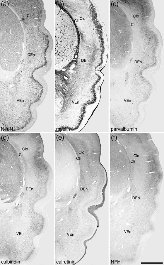
Photomicrographs of coronal sections through the claustrum (Cl) and endopiriform nuclear complex (En) of the tree pangolin stained for neuronal nuclear marker (NeuN, a), myelin (b), parvalbumin (c), calbindin (d), calretinin (e), and neurofilament H (NFH, f). Using this range of stains, the claustrum of the tree pangolin could be divided into inner (Cli) and outer (Clo) divisions, the differentiation of which is most clear in the parvalbumin (c), calbindin (d), calretinin (e), and neurofilament H (f) stained sections, but also apparent in the NeuN (a) and myelin (b) stains. The En could be divided into dorsal (DEn), ventral (VEn) and intermediate nuclei (IEn, not depicted), the distinction between DEn and VEn being primarily based on the calbindin (d) and calretinin (e) immunostaining. In all photomicrographs, medial is to the left and dorsal to the top. Scale bar in (f) = 2 mm and applies to all. See list for abbreviations