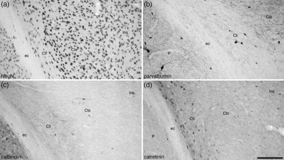
FIGURE 3.

Photomicrographs of coronal sections through the tree pangolin brain showing the architectural appearance of the inner (Cli) and outer (Clo) divisions of the claustrum stained for neuronal nuclear marker (NeuN, a), parvalbumin (b), calbindin (c), and calretinin (d). Note the variation in cellular architecture (a) and the patterns of immunostaining (b‐d) between the inner and outer divisions and the insular cortex (Ins). Also note the absence of an extreme capsule separating the Clo from the Ins, although a clear external capsule (ec) separates the Cli from the putamen (P). In all photomicrographs, medial is to the left and dorsal to the top. Scale bar in (d) = 200 µm and applies to all
