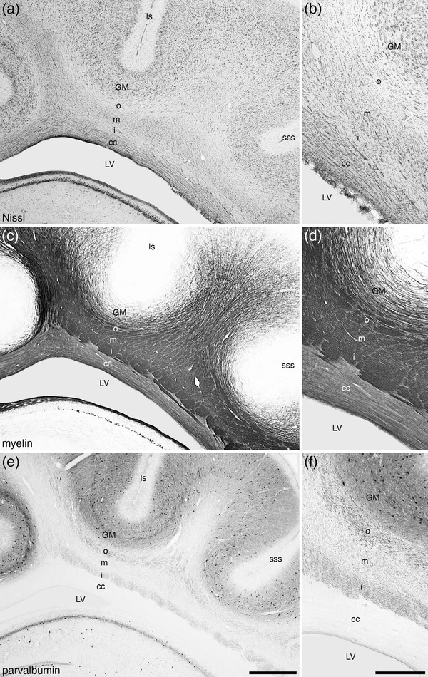
FIGURE 5.

Lower (a, c, e) and higher (b, d, f) magnification photomicrographs of coronal sections through the white matter underlying the cerebral cortex (in this instance deep to the lateral, ls, and suprasylvian, sss, sulci) stained for Nissl (a, b), myelin (c, d), and parvalbumin (e, f) in the brain of the tree pangolin. Note how the cerebral white matter in this species forms three distinct layers, the outer (o), middle (m), and inner (i) layers, here imaged dorsal to the lateral expansion of the corpus callosum (cc) and the body of the lateral ventricle (LV). This lamination of the cerebral white matter, while distinct in both Nissl and myelin‐stained sections, is particularly clear in the parvalbumin‐stained sections (e, f). In all photomicrographs, medial is to the left and dorsal to the top. Scale bar in (e) = 1 mm and applies to a, c, and e. Scale bar in (f) = 500 µm and applies to b, d, and f
