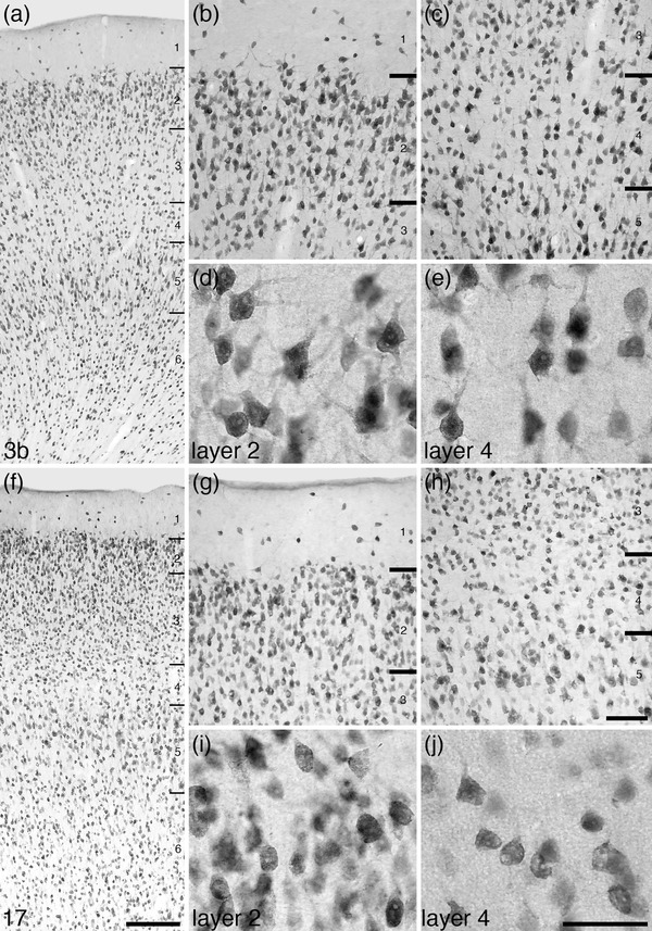
FIGURE 9.

Photomicrographs at different magnifications of neuronal nuclear marker immunostaining coronal sections through the primary somatosensory (Brodmann area 3b, a–e) and primary visual (Brodmann area 17, f–j) cortical areas. In the cortex of the tree pangolin, the neurons of layers 2 and 4 appear to primarily consist of pyramidal neurons (layers labeled in a‐c and f‐h), rather than the typically reported granular neurons (d, e, i, j). Also note that layer 2 exhibits a high neuronal density, while layer 4 is a region of lower neuronal density. These characteristics of the cerebral cortex (only the granular cortex regarding layer 4) are found throughout the cerebral cortex of the tree pangolin. In all images, the pial surface is to the top. Scale bar in f = 250 µm and applies to a, f. Scale bar in h = 100 µm and applies to b, c, g, and h. Scale bar in j = 50 µm and applies to d, e, i, and j
