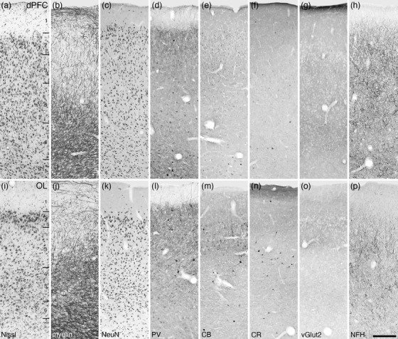
FIGURE 10.

Photomicrographs of the dorsal prefrontal cortical area (dPFC, a‐h) and the lateral orbital prefrontal cortical area (OL, i–p) within the frontal cortex of the tree pangolin stained for Nissl (a, i), myelin (b, j) neuronal nuclear marker (NeuN, c, k), parvalbumin (PV, d, l), calbindin (CB, e, m), calretinin (CR, f, n), vesicular glutamate transporter 2 (g, o), and neurofilament H (NFH, h, p). Both the dPFC and OL lack a distinct layer 4, being agranular cortical areas, while the remaining layers 1–3, 5, and 6, while present (layers labeled in a and i), do not display precise layer boundaries. In all images, the pial surface is to the top. Scale bar in p = 250 µm and applies to all
