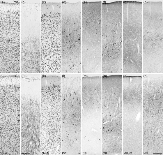
FIGURE 15.

Photomicrographs of the parietoventral somatosensory area (PVS, a‐h) and the second somatosensory area (SII, i–p) of the tree pangolin. Note the low density of neurons in layer 4, and how in both, the upper border of layer 5 is demarcated by the presence of CR‐immunopositive neurons. All conventions, scale bar, and abbreviations as in Figure 10
