
FIGURE 19.
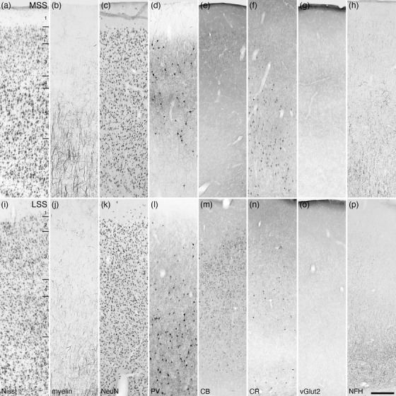
Photomicrographs of the medial (MSS, a‐h) and lateral (LSS, i–p) suprasylvian cortical regions of the tree pangolin. The varied staining patterns of neurons immunopositive for PV, CB, and CR guide the division of this cortical region into the various areas. All conventions, scale bar, and abbreviations as in Figure 10