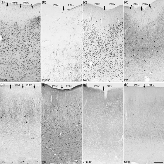
FIGURE 26.

Photomicrographs of the perirhinal cortical areas of the tree pangolin. These agranular five layered cortical areas lie dorsally adjacent to the entorhinal cortex. The perirhinal cortex could be subdivided into dorsal (PRhd) and ventral (PRhv) areas, although there are only subtle differences between these two small cortical areas (the borders of which are marked with arrows), such as variances in the densities of parvalbumin (d) and calbindin (e) immunopositive neurons in layer 3. All conventions, scale bar, and abbreviations as in Figure 10
