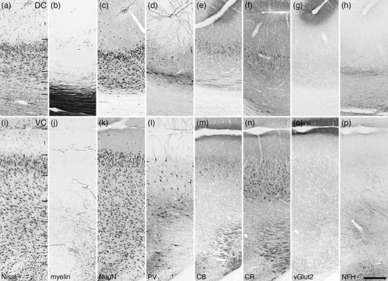
FIGURE 27.

Photomicrographs of the dorsal (DC, a–h) and ventral (VC, i‐p) cingulate cortical areas of the tree pangolin. Both areas lack a layer 4, with the DC being thinner than the VC due to the DC being located around the fundus of the cingulate sulcus. Note that the PV (d, l), CR (f, n), and NFH (h, p) immunostaining is different between the two cortical areas. All conventions, scale bar, and abbreviations as in Figure 10
