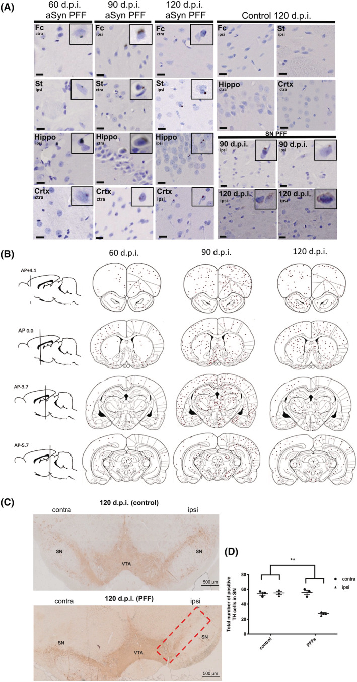
FIGURE 1.

Phosphorylated alpha‐synuclein (aSyn) slowly accumulates in regions distant from the preformed fibril (PFF) injection site. (A) Using a stereological approach, a 1 in 20 sequence of 7 μm coronal sections was taken throughout the entire brain (both contralateral and ipsilateral hemispheres) of animals injected with aSyn PFFs for 60, 90 and 120 d.p.i. or control (vehicle) for 120 d.p.i. Representative images are shown. Sections were immunolabelled with an antibody against aSyn phosphorylated at Ser129 and counterstained with haematoxylin to label nuclei. Phosphorylated aSyn inclusions appeared as faint cytoplasmic and denser perinuclear inclusions in the frontal cortex (Fc), striatum (St), hippocampus (Hippo), cortex (Crtx) and substantia nigra (SN). Control animals did not show any positive labelling. Scale bars = 10 μm, n = 3. (B) The total number of positively immunolabelled cells in three animals from each PFF‐injected group was counted and the positions marked on coronal brain tracings. Cells positive for phosphorylated aSyn were observed in many regions including those directly and indirectly connected to the medial forebrain bundle (MFB) and appeared to peak at 90 d.p.i. The red circle indicates the site of injection (MFB). Regions proximal to the injection site appeared to show the most abundant phosphorylated aSyn positive cells; (C) 7 μm coronal sections from control and PFF‐injected rats at 120 d.p.i. were immunolabelled using an antibody raised against tyrosine hydroxylase (TH). Representative images are shown. Red box identifies region of interest used for quantification. (D) Quantification of TH cell count for 120 d.p.i. tissues. To detect differences in cell number between PFF and control groups, an unpaired t test was used. This showed a statistically significant reduction in TH cell number in the PFF group. **p < 0.01. Data shown are mean ± SEM, n = 3.
