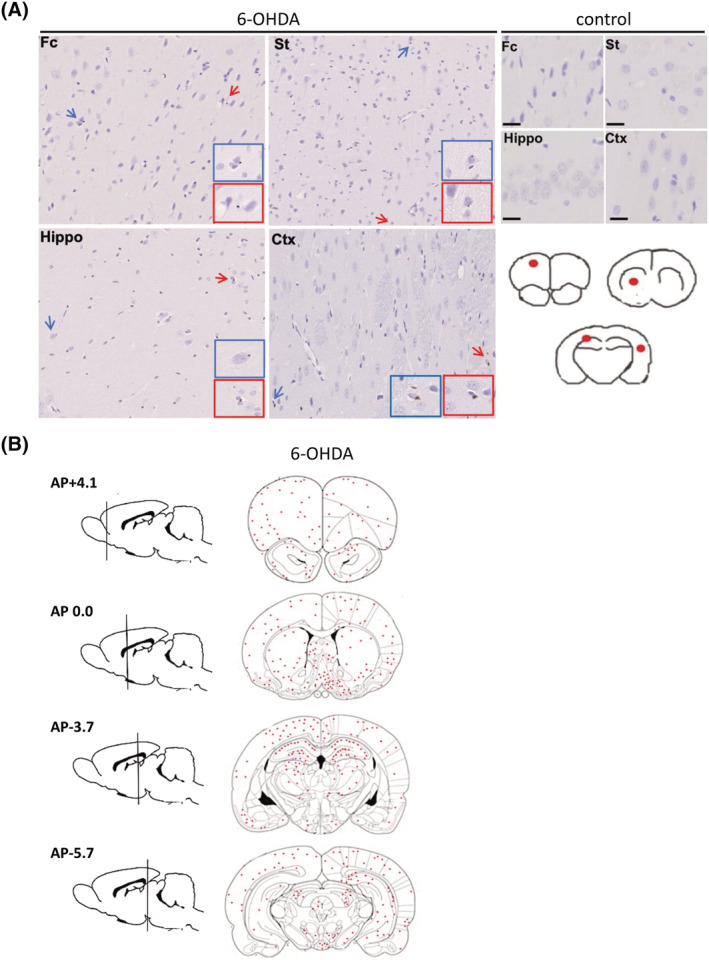
FIGURE 2.

Phosphorylated alpha‐synuclein (aSyn) rapidly accumulates in regions distant from the 6‐OHDA injection site. (A) Immunohistochemistry was performed on 7 μm thick brain tissues from rats unilaterally injected with phosphate‐buffered saline (PBS) (control) or 6‐OHDA into the medial forebrain bundle (MFB). An antibody raised against phosphorylated aSyn at serine 129 (81A) was used to label cells containing phosphorylated aSyn. Control rats (top right panel) did not show any 81A immunoreactivity, whereas cells showed faint positive labelling for phosphorylated aSyn in Fc, St, Hippo and Crtx in response to 6OHDA (middle panel). Red scale bar 20 μm, black scale bar, 5 μm, n = 3. Blue and red arrows indicate cells immunoreactive to the phosphorylated aSyn antibody, with coloured insets showed higher magnification images of these cells. Images are from representative rodents. Fc = frontal cortex, St = striatum, Hippo = hippocampus and Crtx = cortex. Red circles on brain traces (bottom right panel) show the Fc, striatum, hippocampus and cortical areas from which sections were obtained and immunolabelled. (B) The total number of positively immunolabelled cells in three animals from the 6‐OHDA‐injected group was counted and the positions marked on coronal brain tracings. Cells positive for phosphorylated aSyn were observed in many regions including those directly and indirectly connected to the MFB.
