FIGURE 3.

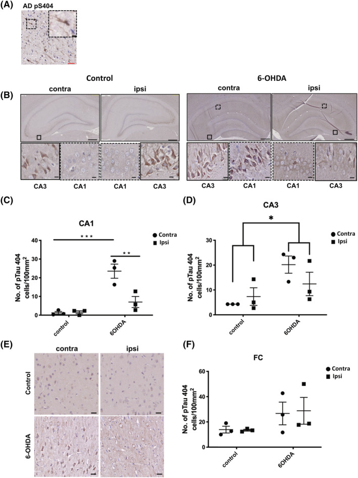
(A) Tau phosphorylated at Ser404 rapidly accumulates in regions distant from the 6‐OHDA injection site. Immunohistochemical staining was performed on 7 μm paraffin embedded sections from the temporal cortex of a post‐mortem Alzheimer's disease (AD) brain using an antibody against pTau404. Inset shows higher magnification of immunoreactive neurons. For rat brain, 7 μm paraffin embedded sections were labelled with the same antibody against pTau404. (B) Representative images of pTau404 immunolabelling at 3 weeks post injection. Sections were counterstained with haematoxylin. Both control and 6‐OHDA‐injected animals showed labelling of pTau404 in CA3. pTau404 labelling was also apparent in CA1 of 6‐OHDA animals but not control animals. Bar charts in show quantification of the number of pTau404 +ve cells in (C) CA1 and (D) CA3. (E) Representative images of pTau404 immunolabelling in frontal cortex (Fc). Sections were counterstained with haematoxylin. Bar chart in (F) shows the number of pTau404 +ve cells in frontal cortex. Scale bars in main images are 500 μm, and 5 μm scale bars are used in insets. Unpaired t tests were used to determine differences in the number of immunoreactive cells between control and 6‐OHDA groups for either the contralateral or ipsilateral hemisphere and demonstrated a statistically significant increase in pTau404‐immunoreactive cells in the CA1 and CA3. No differences were found for the Fc region. *p < 0.05, ***p < 0.001. Data shown are mean ± SEM, n = 3.
