FIGURE 5.

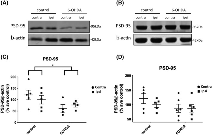
6‐OHDA injection caused a loss of PSD‐95 in the frontal cortex but not hippocampus. (A) Immunoblots of post‐synaptic density protein‐95 (PSD‐95) in the high salt fraction of frontal cortex from 6‐OHDA‐ and control‐injected rats, 3 weeks post‐injection. (A) Representative immunoblots from (A) frontal cortex and (B) hippocampus, probed with an antibody against the post‐synaptic marker, PSD‐95 (95 kDa). β‐Actin was used as a loading control (42 kDa). (C) The dot plot shows a marked reduction in the amount of PSD‐95 relative to β‐actin in frontal cortex of 6‐OHDA‐injected rats relative to controls. Control, n = 5, preformed fibril (PFF), n = 4. (D) The dot plot shows no significant difference as a result of treatment or between hemispheres in the hippocampus, control, n = 5, PFF, n = 6. Unpaired t tests were used to determine differences in the amount of PSD‐95 between control and 6‐OHDA groups for either the contralateral or ipsilateral hemisphere and showed a significant effect of treatment. *p < 0.05. Data are mean ± SEM presented as % average control where control is the ipsilateral hemisphere of control‐injected rats.
