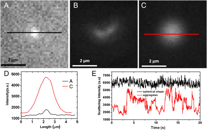
Fig. 4.
Particles imaged by REDFSM in GBS negative clinical samples. (A–C) Snapshots of dark-field scattering images of different types of particles in the GBS-negative samples. (D) Scattering intensity from spherical shape particles in (A) and (C). (E) Scattering intensity curves of particles in (B) (aggregate) and (C) (spherical particle) due to the different shapes.

