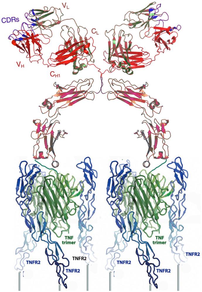
Fig. 1.
A cartoon ribbon diagram depicting the TNFR2 selective agonist cross-linking two sets of three TNFR2s (blue) on a cell surface (posts). The agonist is composed of an IgG1 (red/brown) covalently linked through its carboxyl termini to triplets of a mutated TNFα (green) selective for TNFR2. The cartoon was constructed using data in refs. 12 and 13.

