Fig. 2.
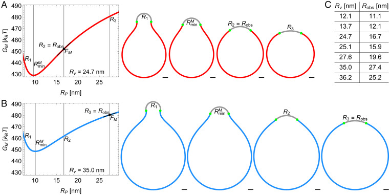
Energy landscape of Piezo vesicle shape. Stationary lipid membrane bending energy as a function of Piezo dome radius of curvature for (A) the Piezo vesicle radius [vesicle 3 in figure 4 of our companion paper (10)] and (B) the Piezo vesicle radius [vesicle 6 in figure 4 of our companion paper (10)] together with selected vesicle cross-sections. These correspond to , in A and in B, , and . In A, is equal to the observed value of , , while in B. For each Piezo vesicle, we describe the Piezo dome geometry as a spherical cap with fixed cap area . The in-plane Piezo dome radius and the Piezo dome contact angle associated with the predicted free membrane shapes in figure 4 of our companion paper (10) determine, for each Piezo vesicle, via . (Scale bars, 5 nm.) (C) The table shows vs. for the seven Piezo vesicles in figure 4 of our companion paper (10). The corresponding spherical cap areas are approximately , , , , , , and (from top to bottom).

