Fig. 2.
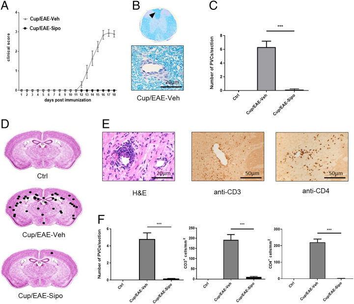
Siponimod ameliorates the disease course and inflammatory demyelination in the Cup/EAE model. (A) Clinical scores for Cup/EAE-vehicle (gray triangles; n = 10) and Cup/EAE-siponimod (black squares; n = 8) groups. (B) Representative LFB/PAS stained section of the spinal cord. Arrowhead indicates an inflammatory PVC shown beneath in higher magnification. (C) Quantification of PVCs in spinal cord sections in control (n = 3), Cup/EAE-vehicle (n = 9) and Cup/EAE-siponimod (n = 8) mice. Differences were determined using one-way ANOVA followed by Tukey's multiple comparisons test. (D) Cumulative spatial distribution of PVCs in the forebrain of control (n = 3), Cup/EAE-vehicle (n = 10), and Cup/EAE-siponimod (n = 8) groups. (E) Histological presentation of a representative forebrain PVC, visualized by H&E stain, anti-CD3, and anti-CD4 immunohistochemistry. (F) Numbers of PVCs in the entire brain as well as CD3+ and CD4+ lymphocyte densities in the corpus callosum (CC). Differences were determined using one-way ANOVA followed by Tukey's multiple comparisons test. Data are shown as mean ± SEM. ***P < .001.

