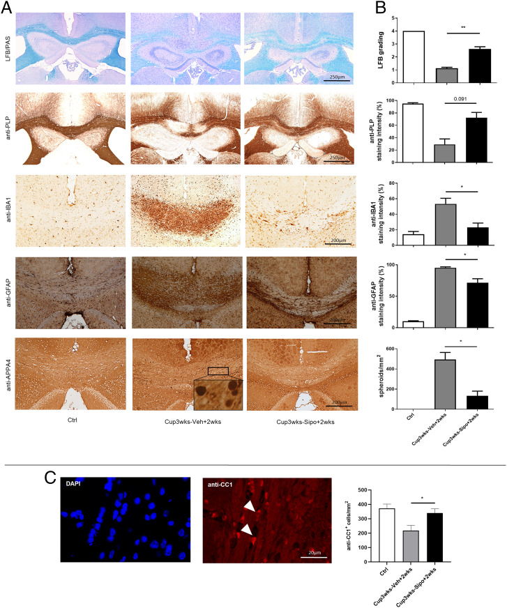
Fig. 3.
Siponimod ameliorates cuprizone-induced pathologies. (A) Representative LFB/PAS, anti-PLP, anti-IBA1, anti-GFAP and anti-APP A4 stains of the medial CC from control (n = 3), Cup3wks-Veh+2wks (n = 8) and Cup3wks-Sipo+2wks (n = 9) treated cuprizone-intoxicated mice. (B) Evaluation of the extent of demyelination (LFB/PAS and anti-PLP), microgliosis (anti-IBA1), astrocytosis (anti-GFAP) and acute axonal injury (anti-APP A4). Statistical analyzes were performed by Kruskal–Wallis test followed by Dunn’s multiple comparison test (C) Representative anti-CC1 immunofluorescence staining of the medial CC. Arrowheads indicate the localization of anti-CC1+ cells. (D) Quantification of CC1+ cell numbers. Statistical analyzes were performed by Kruskal–Wallis test followed by Dunn’s multiple comparison test. Data are shown as mean ± SEM. *P < 0.05; **P < 0.01.

