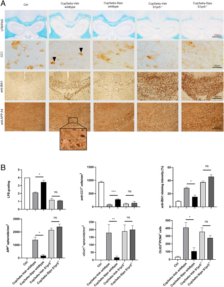
Fig. 8.
Protective siponimod effects are absent in S1pr5−/− mice. (A) Representative LFB/PAS, anti-CC1, anti-IBA1 and anti-APP A4 stains of the medial CC from control (n = 4), Cup3wks-Veh-wildtype (n = 4), Cup3wks-Sipo-wildtype (n = 4), Cup3wks-Veh-S1pr5−/− (n = 5) and Cup3wks-Sipo-S1pr5−/− (n = 5) mice. Arrowheads highlight CC1+ mature oligodendrocytes. (B) Evaluation of the histological results of one experiment. The results have been verified in a second independent experiment (i.e., absence of protective siponimod effects in S1pr5−/− mice; see SI Appendix, Fig. S6). Statistical analyzes were performed by two separate Mann–Whitney tests as indicated. Data are shown as mean ± SEM. *P < 0.05; **P < 0.01; ***P < 0.001. ns, nonsignificant.

