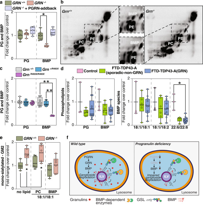
Fig. 4. BMP levels are reduced in progranulin-deficient cells or brain tissues.
a Quantification of PG and BMP isolated from GRN+/+ (green), GRN−/− (orange), and GRN−/− + PGRN-addback (blue) HeLa cell lines (n = 7) reveals a ~50% reduction in BMP levels in PGRN-knockout cells. b Labeling of cellular lipids by feeding 14C-arachidonic acid–albumin complex to GRN+/+ and GRN−/− HeLa cell lines (60 min). Inset highlights reduced levels of BMP in Grn−/− HeLa cells, compared to control cells. c Quantification of PG isolated from Grn+/+ (gray) (n = 8), Grn+/R493X (blue) (n = 8), and GrnR493X/R493X (purple) (n = 8) and quantification of BMP isolated from Grn+/+ (gray) (n = 8), Grn+/R493X (blue) (n = 6), and GrnR493X/R493X (purple) (n = 6) mouse brains reveals a decrease in BMP levels upon loss of PGRN in mouse brains (~50%). d Quantification of PG and BMP isolated from the frontal lobes of control (pink) (n = 3), FTD-TDP43-A(sporadic-non-GRN) (green) (n = 6), and FTD-TDP43-A(GRN) (blue) (n = 11) human brains. BMP species with mono- or di-unsaturated fatty acid moieties are not different, whereas BMP species containing two docosahexanoic acid moieties (22:6/22:6) are reduced in the frontal and occipital lobes of all FTD subjects. e Quantification of GM2 species isolated from Grn +/+ (green) (n = 6) and Grn−/− (orange) (n = 6) HeLa cells after feeding no lipids, di-oleoyl PC or di-oleoyl BMP. f Model of the role of progranulin in the degradation of gangliosides. PGRN/granulin deficiency leads to reduced BMP levels through unclear mechanisms. Reduced BMP levels contribute to impaired ganglioside degradation. Eventually, this leads to lysosomal dysfunction and downstream consequences, including neuroinflammation and neurodegeneration. Box plots display mean ± the minimum and maximum number. One-way ANOVA, followed by multigroup comparison (Dunn’s) test, was performed. *p < 0.05, **p < 0.01.

