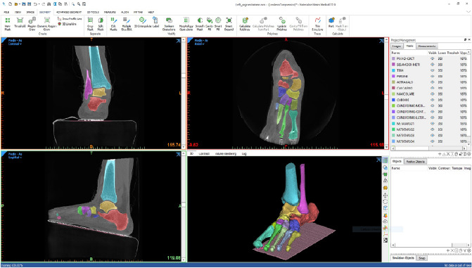
Figure 1.
A screenshot from the 3D reconstruction of the foot bones from a CBCT scan by Mimics Innovation Suite (Materialise, Belgium) software. Each bone segment has a different colour, in both the 3D view (bottom-right) and in the three anatomical planar views; in this antero-posterior slice (top-left), the tibia, fibula, calcaneus and talus bone segments are depicted. In the sagittal view (bottom-left), apparently the foot is an inclined ground, but this is accounted for to the inclination of the gantry in the present device, i.e. the foot is on the horizontal ground.

