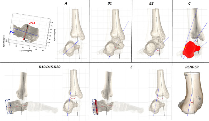
Figure 2.
Representation of all the techniques for HAA calculation. In the frontal view, the longitudinal axis of the tibial distal diaphysis (black) and the hindfoot vertical axis (blue) are shown for each technique. For technique A, the three principal component axes of the PCA applied to the calcaneus are also shown. For techniques D and E, the sagittal views are also to better represent the posterior part of the calcaneus. Lastly, the bottom-right (RENDER) is the posterior view of the CBCT rendering of the same scan.

