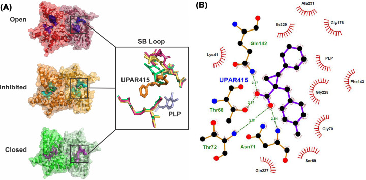
Figure 8. Inhibition of StCysK by UPAR415.
(A) Structural comparison of the substrate binding loop between the open conformation (1OAS) shown in shades of red, the inhibited conformation (6Z4N) shown in shades of orange, and the closed conformation (1D6S) shown in shades of green. (B) LigPlot showing the interactions between the enzyme residues and the UPAR415 molecule. Figure produced using PyMOl and LigPlot.

