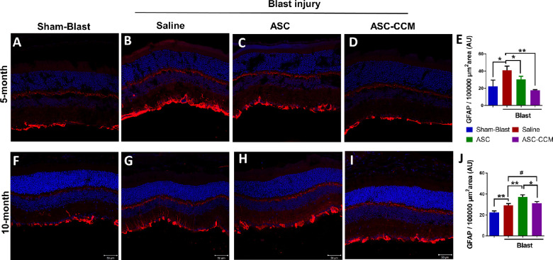
Figure 4.
ASC-CCM and ASCs in mice subjected to blast injury partially suppress glial hypertrophy. Confocal microscope images of retinal tissue immunolabeled for GFAP in (A) sham mice receiving saline solution, five months; (B) mTBI mice receiving saline solution, five months; (C) mTBI mice receiving ASCs, five months; (D) mTBI mice receiving ASC-CCM, five months. (E) Image J quantification of GFAP intensity in immunolabeled retinas at five months. (F) Sham mice receiving saline solution, 10 months. (G) mTBI mice receiving saline solution, 10 months. (H) mTBI mice receiving ASCs, 10 months. (I) mTBI mice receiving ASC-CCM, 10 months. (J) Image J quantification of GFAP intensity in immunolabeled retinas at 10 months. Scale bars for A-I: 50 µm. Data represent mean ± SEM from n = 5-8 animals/group. *P < 0.05; **P < 0.01; #P > 0.05.

