
Figure 5.
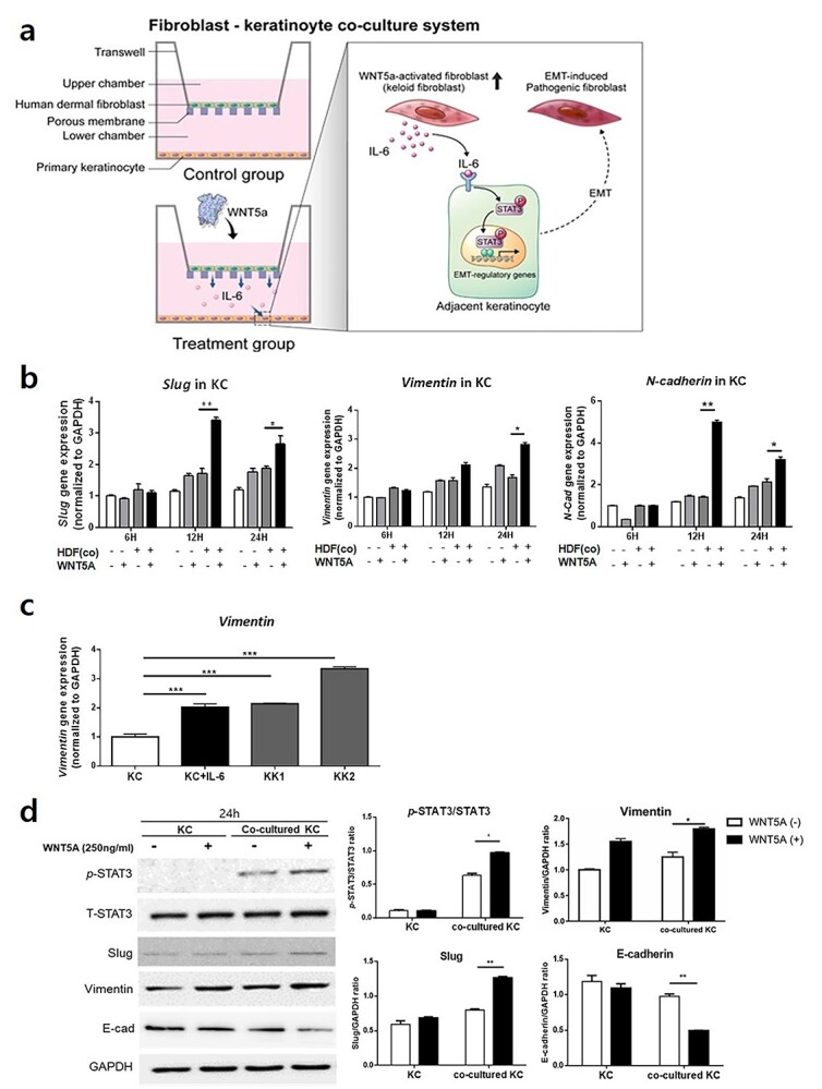
KCs express elevated levels of EMT markers during co-culture with WNT5A-activated HDFs or IL-6 stimulation. (a) Illustration of the co-culture system involving KCs and HDFs. (b) Increased levels of slug, vimentin and N-cadherin following co-culture of KCs with WNT5A-activated HDFs. (c) IL-6-stimulated KCs as well as patient-derived keloid keratinocytes from two different keloid tissues (KK1, KK2) show elevated gene expression levels of vimentin compared to the normal keratinocytes. (d) Western blot analysis of KCs co-cultured for 24 h with WNT5A-treated HDFs shows significantly increased levels of p-STAT3, slug and vimentin and decreased level of E-cadherin. Data are from three independent experiments. *p < 0.05, **p < 0.01, ***p < 0.005; Mann–Whitney U test. KC keratinocytes, KK keloid keratinocytes, HDF human dermal fibroblasts, EMT epithelial-mesenchymal transitio, WNT5A Wnt family member 5A, IL interleukin