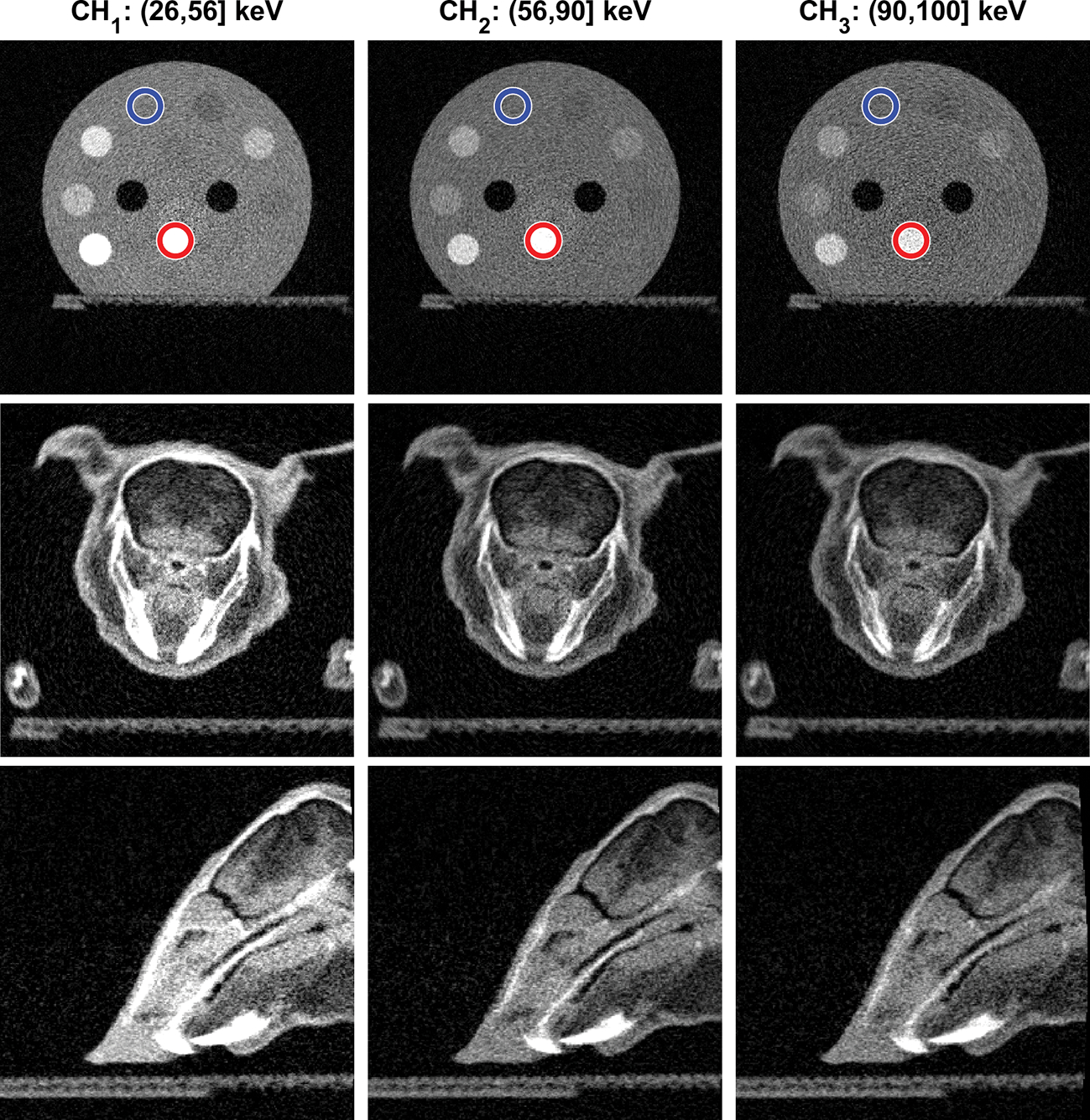
Figure 6:

Top row: CBCT images of the CT calibration insert phantom in the three energy channels. Blue and red circles indicate the regions of water and bone basis materials used in material decomposition. Middle and Bottom rows: CBCT images of the plastinated mouse phantom on transverse and sagittal planes respectively. Display window [0,0.5] cm−1 for all images.
